Capillary electrophoresis (CE) is a versatile and increasingly essential tool for analyzing a broad spectrum of analytes, from small inorganic ions to large DNA macromolecules. Offering reliable data with minimal sample preparation and a high degree of automation, CE serves as a valuable alternative to traditional methods like gel electrophoresis and liquid chromatography. This technique effectively detects both high- and low-affinity molecular interactions, separating charged and neutral molecules with ease. Through various CE modes, separation is achieved based on charge, size, and frictional forces, allowing for rapid, efficient separations in a range of fields.
A Growing Role across Analytical Fields
CE is now a well-established technique in analytical labs, capable of detecting binding interactions from millimolar to nanomolar concentrations. It is particularly beneficial in drug discovery and screening, where it aids in the analysis of enzymes, receptor domains, structural proteins, nucleic acid complexes, bioactive peptides, protein-protein interactions, and antibodies. This wide applicability underscores CE as a proven, powerful technology in multiple domains.
Instrumentation: The Core Components of CE
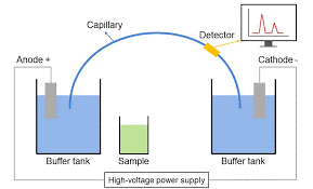
A typical capillary electrophoresis system includes:
- High-voltage power supply: Initiates analyte separation by applying an electric field.
- Sample introduction mechanism: Controls sample entry into the capillary tube.
- Capillary tube: Usually made of fused silica, sometimes polyimide-coated, and filled with an electrolytic buffer.
- Detector and output device: Records results based on analyte absorption and generates an analyte mass spectrum for characterization.
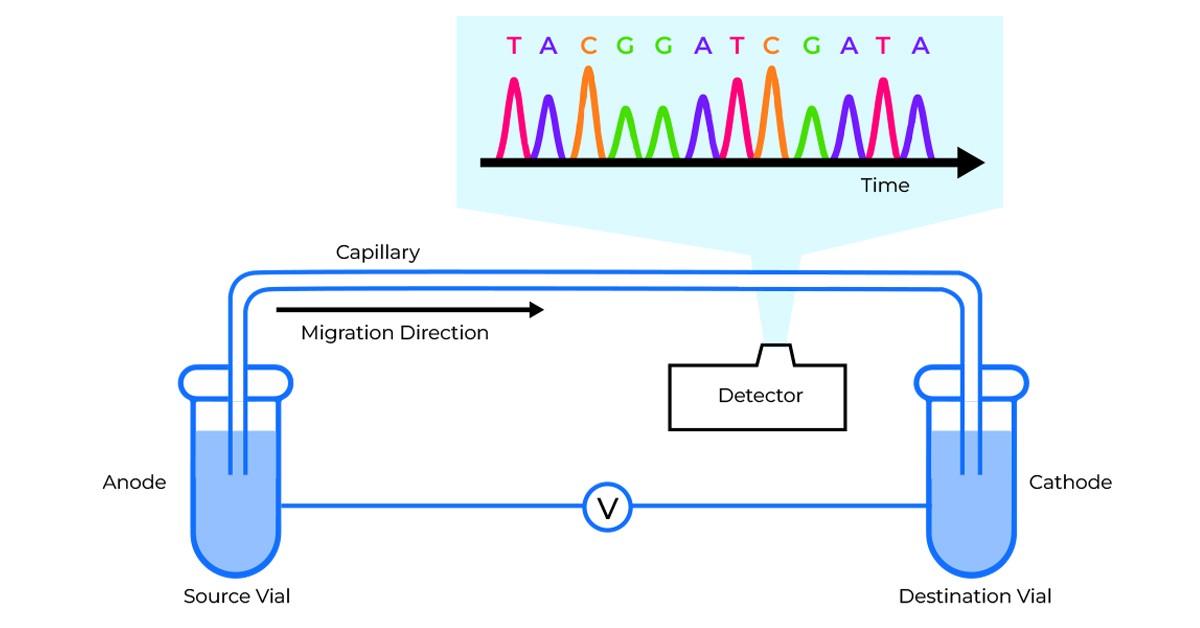
For added precision, some systems incorporate a temperature control unit to stabilize the separation, as temperature influences electrophoretic mobility. The detector, typically positioned near the tube’s cathodic end, passes UV-VIS light through a small window for absorbance measurement, while a photomultiplier tube provides further details on mass-to-charge ratios of ionic species.

Figure 1: Schematic diagram of a typical capillary electrophoresis workflow
Mechanisms of Separation: Electrophoretic Mobility and Electroosmotic Flow
In capillary electrophoresis, analyte separation relies on two main mechanisms:
- Electrophoretic mobility: Drives charged particles through the capillary based on their charge-to-mass ratio.
- Electroosmotic flow (EOF): Moves buffer ions within the electric field, intersecting with electrophoretic migration to enhance separation. EOF’s flow profile can be fine-tuned via electrolyte pH and capillary surface charge, which reduces band broadening and delivers sharper peaks than HPLC.
Various detection systems (UV-Vis, PDA, laser-induced fluorescence) quantify analytes, offering similar advantages to those of HPLC.
Modes of Capillary Electrophoresis
- Capillary Zone Electrophoresis (CZE): The most common CE mode, ideal for both small and large molecules. Analytes migrate at speeds based on charge differences, forming distinct zones. Adding chiral selectors to CZE enables the analysis of enantiomers, useful for tasks like D- and L-amino acid separation.
- Capillary Gel Electrophoresis (CGE): Used for macromolecules like proteins and oligonucleotides. Here, a gel-filled capillary acts as a molecular sieve, providing size-based separation and facilitating the characterization of biopharmaceutical macromolecules.
- Capillary Isoelectric Focusing (CIEF): Ideal for molecules with unique isoelectric points (pI). CIEF’s pH gradient separates molecules by pI, which halts migration when pI is reached. It’s particularly effective for analyzing mAbs and other therapeutics with natural heterogeneity.
Advantages of CE over HPLC
CE offers significant advantages over high-performance liquid chromatography (HPLC):
- Higher separation efficiency and shorter analysis times
- Low sample volumes and chiral separation without costly columns
- Versatility and compatibility with various detection systems
Additionally, CE’s reliance on aqueous solutions rather than organic solvents makes it an environmentally friendly choice. Unlike HPLC, CE’s separation mode depends only on the buffer, allowing a single capillary to support multiple modes of separation. This adaptability has contributed to its acceptance across diverse analytical settings.

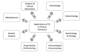
Figure 2: Applications of CE in Clinical Chemistry
Next-Generation Sequencing (NGS) and Capillary Electrophoresis (CE): A Comparison
Next-Generation Sequencing (NGS) has emerged as an alternative and complement to CE, particularly in forensic DNA analysis. Here’s how they compare:
Capillary Electrophoresis (CE)
- Primarily for STR profiling: CE remains highly effective in detecting Short Tandem Repeats (STRs), the cornerstone of forensic DNA analysis.
- Established technology: CE is the gold standard for forensics, supported by well-defined protocols and legal recognition.
- Cost-effective: CE is affordable for routine forensic testing, especially in cases requiring STR analysis.
Next-Generation Sequencing (NGS)
- Provides comprehensive data: NGS sequences entire genomes or targeted regions, offering more insights into ancestry, traits, and microbial DNA.
- Simultaneous multi-marker analysis: NGS can analyze STRs, SNPs, and other markers in one run, making it more versatile.
- Higher sensitivity: NGS can detect low-level DNA and resolve mixed samples, benefiting complex cases.
Challenges with Replacing CE with NGS
- Cost and infrastructure: NGS systems are expensive, and labs require specialized infrastructure.
- Data complexity: NGS generates large data volumes, necessitating advanced bioinformatics expertise.
- Forensic standards: CE’s reliability makes it the legal standard, while NGS is gradually gaining acceptance.
In forensic workflows, CE is still preferred for routine STR analysis, while NGS is often reserved for specialized cases where high sensitivity and more genetic information are critical.
A Promising Future
Capillary electrophoresis is solidifying its role in biopharmaceutical quality control and characterization, especially for therapeutic proteins and oligonucleotides. With its high sensitivity and versatility, CE is comparable to HPLC, with a growing presence in various labs. As NGS adoption increases, CE and NGS together are providing complementary strengths to forensic workflows, ensuring that both technologies will continue shaping the future of genetic analysis.


